We’re Not Just Showing Off. See How Dental Tech Benefits You.
Diagnostics
These diagnostic tools are designed to help us detect issues early and plan treatments with greater accuracy. This could mean more effective care with fewer surprises along the way.
- Digital X-rays: Capture high-quality images with up to 90% less radiation exposure compared to traditional X-rays, enabling immediate and accurate assessments.
- Digital Panoramic & Cephalometric X-rays: Provide comprehensive views of your oral structures, aiding in precise treatment planning for orthodontics and other procedures.
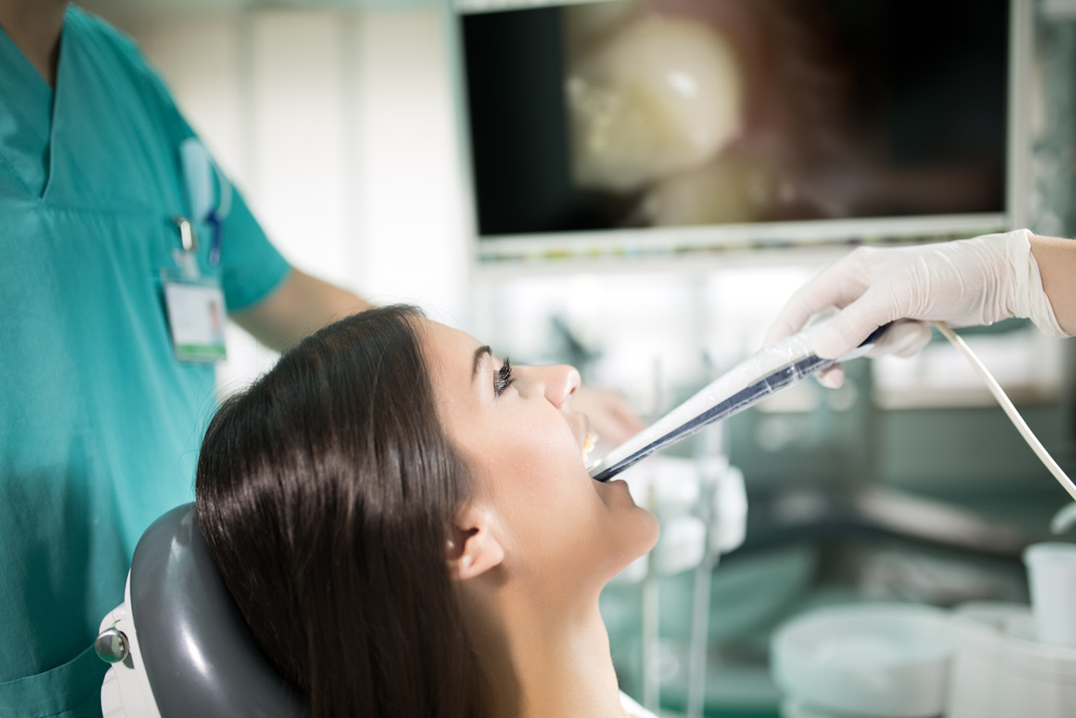
- Intraoral Cameras: Offer real-time, magnified images of your mouth, helping you see what we see and understand your oral health better.
- CariVu™ Cavity Detection: Uses transillumination technology to identify cavities and cracks without radiation, supporting early and safe detection.
- iTero® Digital Impressions: Create precise 3D models of your teeth and gums, eliminating the need for messy traditional impressions.
- Cone Beam Computed Tomography (CBCT): Generates detailed 3D images of dental structures, soft tissues, and nerve paths, facilitating accurate diagnoses and treatment plans.

Orthodontics
These cool technologies are designed to straighten your teeth, monitor your progress, and adjust treatments as needed. Know what else is cool? We’re accepting new orthodontic patients!
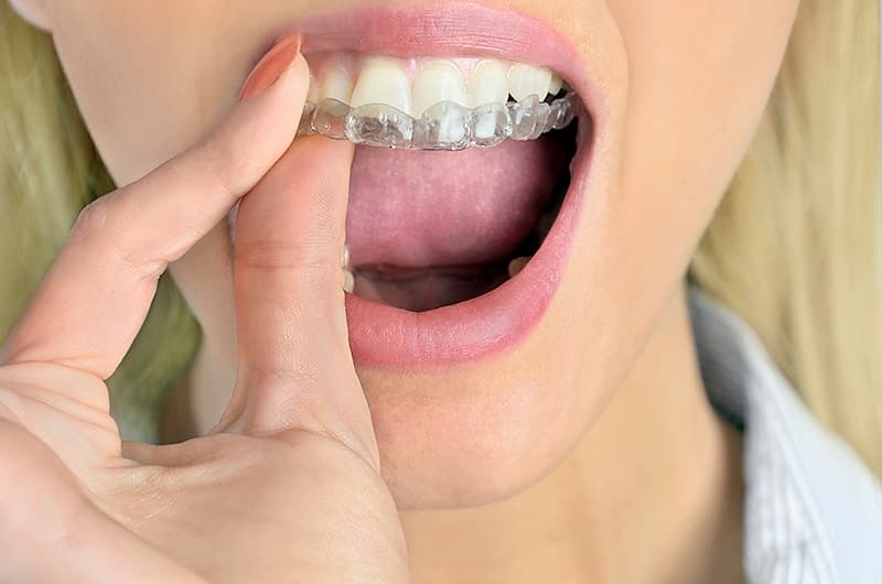
- Spark™ Clear Aligners: Utilize TruGEN™ material for enhanced clarity and comfort, effectively guiding your teeth into alignment.
- Invisalign® and Invisalign Teen®: A discreet way to straighten teeth, with custom-made clear aligners that fit snugly and gradually shift teeth into place.
- Damon™ Smile Braces: Feature a self-ligating system designed to reduce friction and allow for quicker, more comfortable tooth movement.
- Propel® Accelerated Orthodontics: Employs advanced technology to stimulate bone remodeling, potentially reducing treatment time.
- Dental Monitoring: An AI-powered system that enables virtual check-ins, allowing us to track your progress remotely and adjust treatments as necessary.

Hygiene & Gum Disease Treatment
Advanced dental technology helps us watch for, treat, and even prevent gum disease, giving your gums a great fighting chance to stay healthy.
- Guided Biofilm Therapy (GBT) with EMS Airflow®: Combines air, water, and powder to gently remove plaque and biofilm, enhancing gum health and comfort. Great for after braces!
- LANAP® (Laser-Assisted New Attachment Procedure): A minimally invasive laser treatment that targets diseased gum tissue while preserving healthy tissue, promoting natural healing.
- LAPIP® (Laser-Assisted Peri-Implantitis Procedure): Similar to LANAP, this laser therapy treats inflammation around dental implants, aiding in their longevity.
- OralDNA® Saliva Testing: Analyzes saliva to detect harmful bacteria and genetic markers, allowing for personalized periodontal treatment plans.
- Overjet Dental AI: An artificial intelligence tool that interprets dental X-rays, highlighting areas of concern such as bone loss and gum inflammation for early intervention.
TMD/TMJ Treatment Technology
This specialized technology helps diagnose and treat temporomandibular joint disorders (yeah, it’s a mouthful), aiming to alleviate pain and restore function.
- Kois Deprogrammer: A removable appliance used to relax jaw muscles and identify the optimal bite position, aiding in the diagnosis and treatment of TMD.

Don’t Miss Out on Modern Technology
We’d love to show you our advanced tools and how they can enhance your experience. Give us a call!
